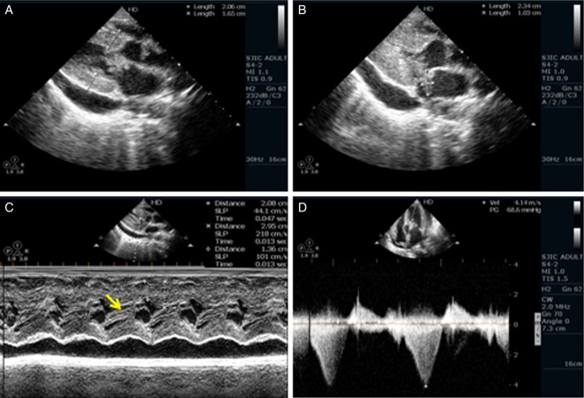
Figure 1.
(A) Parasternal long axis (PLAX) view of septal hypertrophy and a vegetation on the anterior mitral leaflet (AML). (B) PLAX view of a large vegetation on AML and pericardial effusion. (C) M-mode echocardiography showing systolic anterior motion of AML. (D) Continuous wave Doppler showing significant left ventricular outflow gradient of 68 mm Hg.

