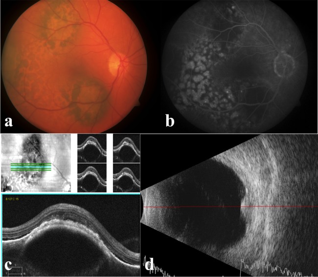
Figure 1.
Manifestations of bilateral diffuse uveal melanocytic proliferation at initial presentation: (A) fundus photo showing flat to minimally raised pigmented choroidal lesions, each was 4–5 mm in basal diameter, with orange-brown, giraffe-skin-like oval patches; (B) fluorescein angiography showing areas of hypofluorescence corresponding to the pigmented choroidal tumours. Hyperfluorescent spots alternating with reticular areas of blocked fluorescence; (C) optical coherence tomography showing the pigmented lesions as areas of focal choroidal thickening, with overlying areas of retinal pigment epithelium atrophy and depositions; (D) ultrasonography showing the pigmented lesions as multiple focal choroidal thickenings with medium internal reflectivity and focal retinal detachments.

