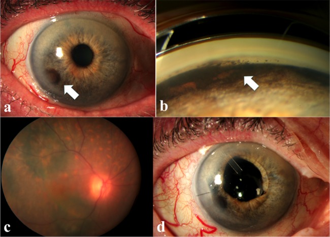
Figure 2.

Progression of bilateral diffuse uveal melanocytic proliferation after 50 months of initial presentation: (A) anterior segment photo showing growth of new iris-pigmented masses after plasma exchange; (B) increased pigment deposition in the angle; (C) increase in the dimensions of the pigmented choroidal lesions; (D) appearance of the eye after phacoemulsification and Ahmed valve surgeries.
