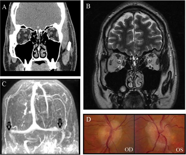
Figure 3.
A. Coronal view of CT of the head without contrast showing dehiscence of the right cribiform plate (asterisk) with soft tissue in the right olfactory recess, consistent with meningocele in this region; B. T2 coronal view of brain MRI showing hyperintense fluid and soft tissue in the right olfactory recess (asterisk) and distension of the peri-optic nerve subarachnoid CSF space (white arrows); C.Head MRV showing bilateral distal transverse stenoses (black arrows). D. Bilateral chronic papilledema.

