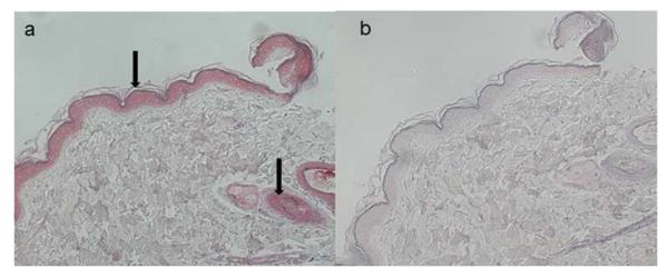
Fig. 1. Simian varicella virus (SVV) antigen in skin rash during reactivation.

SVV antigens are seen in the epidermis and surrounding hair follicles after staining with rabbit anti-SVV gH and gL antibodies (a, arrows), but not in adjacent sections stained with normal rabbit serum (b).
