Fig. 1.
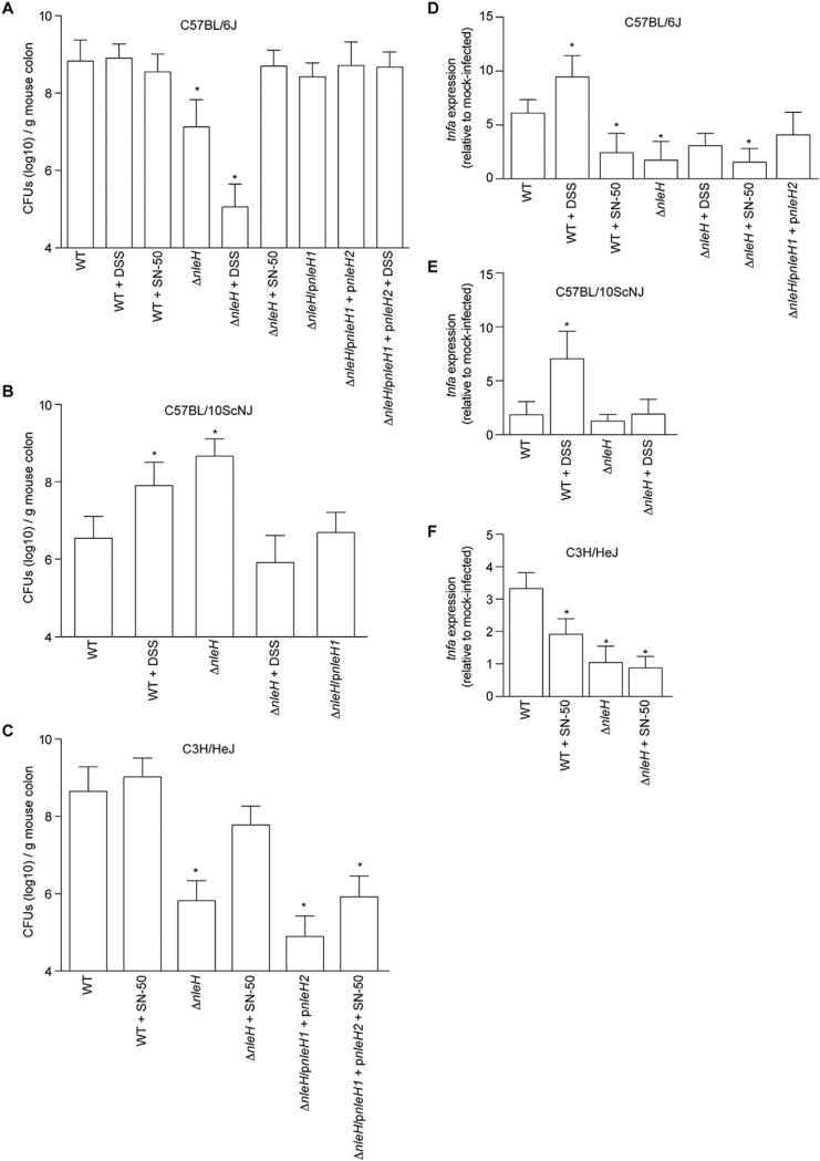
The contribution of NleH to C. rodentium colonization depends upon the mouse strain. A. C. rodentium colonization of C57BL/6J mice. Colonization (CFUs/g mouse colon) of indicated C. rodentium strains in C57BL/6J mice on days 7–11 post-infection. Where indicated, mice were treated with 2.5% (w/v) dextran sodium sulfate (DSS) for 7 d prior to infection or were injected with 10 μg/kg SN-50 immediately prior to gavage. B. C. rodentium colonization of C57BL/10ScNJ mice on days 7–11 post-infection. C. C. rodentium colonization of C3H/HeJ mice. In all panels, asterisks indicate significantly different colonization magnitude (Kruskal–Wallis test; n = 7–15/group; p < 0.05) as compared with WT C. rodentium on days 1–7 post-infection. D–F. TNF expression. Relative TNF mRNA expression levels from colons obtained from mice (7 days post-gavage) infected with the indicated C. rodentium strains. Asterisks indicate significantly different TNF expression as compared with WT infection (t-test; n = 6; p < 0.05).

