Figure 1.
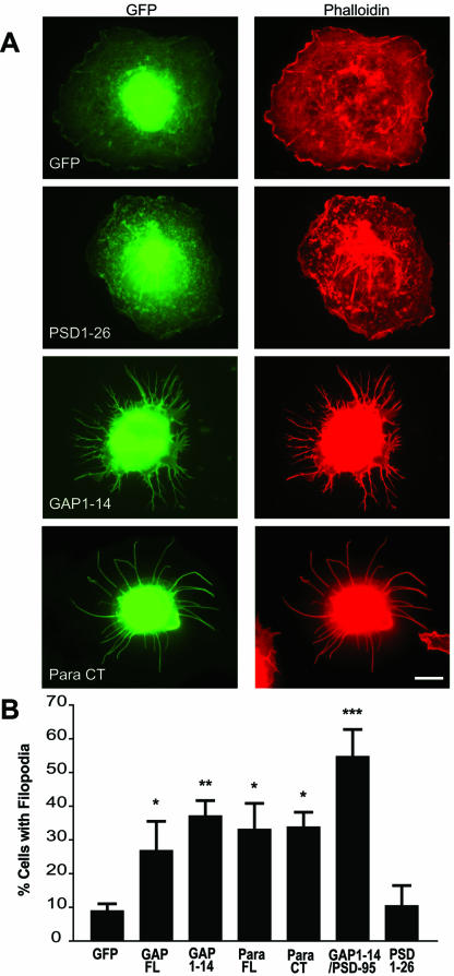
Filopodia induction in COS-7 cells. (A) COS-7 cells were transiently transfected with various constructs fused to GFP (green) as described in Table 1 and immunolabeled for F-actin with a rhodamine-conjugated phalloidin antibody (red). COS-7 cells transfected with full-length GAP-43 (GAP FL) and full-length paralemmin (Para FL) show increased branching vs. control GFP-transfected cells. When transfected with palmitoylation motifs of GAP-43 (GAP1–14) or of paralemmin (Para CT), COS-7 cells show extensive filopodial outgrowth, compared with cells transfected with the palmitoyaltion motif of PSD-95-GFP (PSD1–26). Filopodia induction was quantified by counting the number of cells showing extensive filopodia outgrowth (at least 5 filopodia of ≥10 μm per cell, or at least 20 filopodia of ≥5 μm) and expressed as a percent of cells “with filopodia.” (B) A graph showing that filopodial induction by GAP1–14, Para FL, Para CT, and GAP1–14/PSD-95 are statistically different from GFP (*p < 0.05; **p < 0.01; ***p < 0.01). Scale bar, 10 μm.

