Fig. 2.
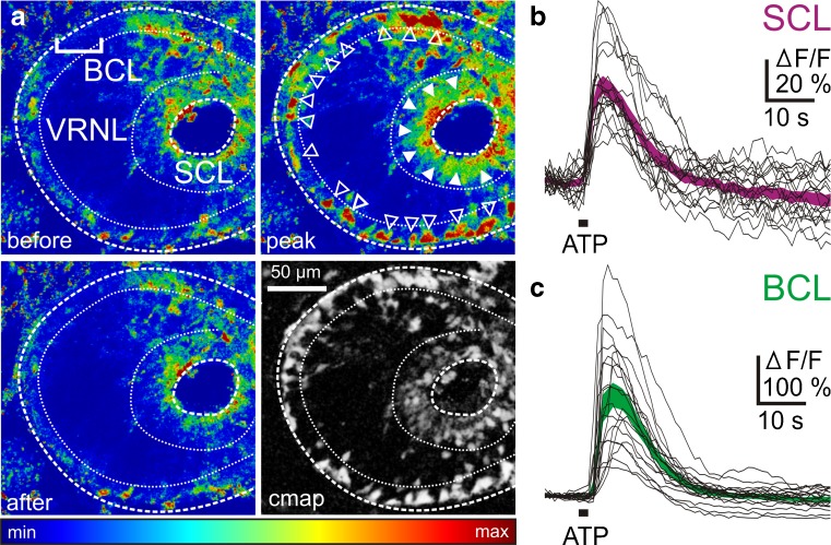
ATP-induced [Ca2+]i increases in supporting and basal cells of the epithelium of the VNO of larval Xenopus laevis. a Pseudocolored image of an acute VNO slice stained with the Ca2+ indicator dye Fluo-4 (SCL supporting cell layer; VRNL vomeronasal receptor neuron layer; BCL basal cell layer). The upper left-hand image was acquired before application of ATP. Application of ATP-induced [Ca2+]i transients in cells of the BCL (open arrowheads) and SCL (filled arrowheads). No apparent changes in Ca2+-dependent fluorescence in cells of the VRNL (upper right-hand image). The lower left-hand image was taken after return to the base line fluorescence. A pixel correlation map (see Materials and methods for details) of the same slice is depicted in the lower right-hand image. Responsive cells appear bright on dark background. b ATP-induced [Ca2+]i transients of individual cells from the SCL (black traces). The magenta-colored area gives the mean [Ca2+]i transients ± SEM of all individual SCs (n = 20). c ATP-induced [Ca2+]i transients of individual cells from the BCL (black traces). The green-colored area gives the mean [Ca2+]i transients ± SEM of all individual BCs (n = 20)

