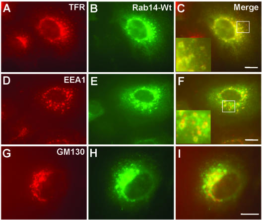
Figure 5.
GFP-Rab14wt fusion protein is primarily localized to TfR-positive compartments. After PFA fixation and saponin permeabilization, GFP-Rab14 wt cells were stained with mouse monoclonal antibodies, TfR (A), EEA1 (D), and GM130 (G), followed by incubation with Alexa-594–labeled anti-mouse IgG antibody. Green corresponds to the GFP signal (B, E, and H). (C, F, and I) Merged images. Note that GFP-Rab14 colocalizes with TfR much better than with EEA1. Partial colocalization of GFP-Rab14 is seen with the Golgi marker GM130. Bars, 10 μm.

