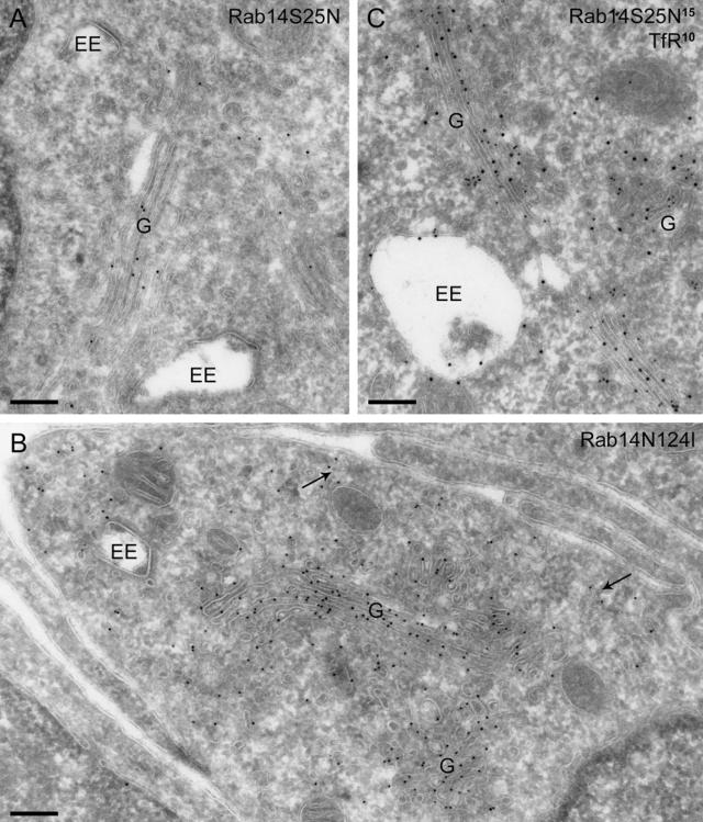
Figure 9.
Inactive Rab14 shifts the labeling pattern toward the Golgi complex. (A) GFP-Rab14S25N, labeled with anti-GFP (10-nm gold), is mainly present on the Golgi complex (G) and largely absent from tubulo-vesicular structures near EEs. (B) Similar distribution is observed in cells transfected with GFP-Rab14N124I (10-nm gold). Some label is also present on small, dispersed vesicles (arrows). (C) Double labeling of GFP-Rab14S25N and TfR shows that the only significant overlap is in the Golgi complex, with virtually no colocalization in tubules or vesicles near endosomes. Bars, 200 nm.

