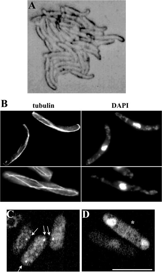
Figure 3.
Gfh1p localizes to the SPB and the EMTOC and its overexpression results in polarity and microtubule defects. (A) Cells of a microcolony overexpressing gfh1+. (B) Cells overexpressing gfh1+ were fixed and stained with either TAT-1 antibodies to visualize microtubules (tubulin) or DAPI to visualize DNA. (C and D) Cells expressing Gfh1p-GFP were subjected to live imaging using a GFP filter set. The localization of the fusion protein to SPBs is indicated by arrows (C) and to the medial EMTOC ring structure in a binucleate cell is indicated by an asterisk (D). Scale bar, 10 μm.

