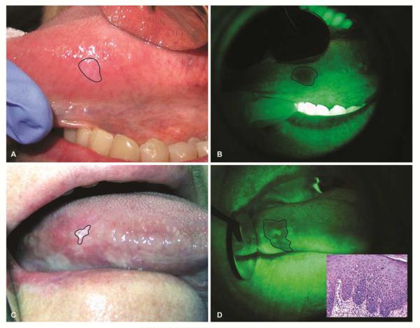
Figure 1.
Autofluorescence visualization of tongue leukoplakias. (A) A 57-year old female with a history of cigarette smoking presented with a leukoplakia that is barely visible under white light. (B) Autofluorescence visualization revealed loss of fluorescence of this leukoplakia. Excisional biopsy of this leukoplakia revealed moderate epithelial dysplasia. (C) A 65-year old female with no history of tobacco use presented with a leukoplakia in her lateral surface of the tongue. Extent of the leukoplakia involvement is markedly different when examined under white light (C) compared to autofluorescence visualization (D). Incisional biopsy of the lesion revealed moderate epithelial dysplasia (Inset).

