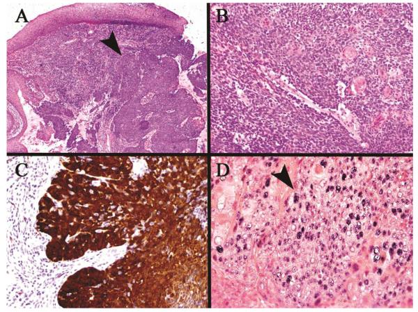
Figure 6.
HPV associated squamous cell carcinoma. (A) Low power view of the tumor arising in the tonsillar cryptic mucosa (arrow) deep to the surface mucosa, (B) Higher power of monotonous, non-keratinizing neoplastic cells typical of this phenotype, (C) Strong/over-expressed p16 immunohistochemical staining throughout the tumor, (D) HPV positive in situ hybridization staining of the tumor nuclei (arrow)

