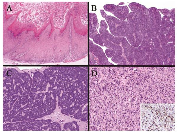
Figure 7.
Histologic variants of squamous cell carcinoma. (A)Verrucous carcinoma showing a broad base, minimal cytologic atypia and exophytic spire growth;(B) Papillary squamous carcinoma showing exophytic growth of fibrovascular cores covered by full thickness neoplastic cells without keratinization; (C) Basaloid squamous carcinoma with high-grade features and scant cytoplasm often lacking keratinization as in this example; (D) Sarcomatoid (spindle cell) carcinoma haphazardly growing in sheets with pleomorphism and mitoses, often retaining cytokeratin expression detected by immunohistochemical staining (inset) which aids in differentiating from true sarcomas.

