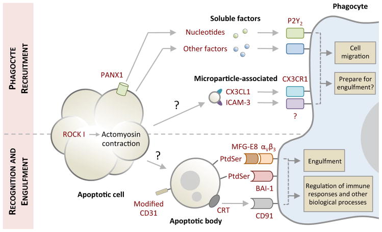
Figure 1. Phases of apoptotic cell clearance.
Cells undergoing apoptosis often exhibit morphological changes (for example membrane blebbing and cellular shrinkage) to facilitate cell detachment and organelle fragmentation. Prior to or during the onset of apoptotic morphology, apoptotic cells also release ‘find-me’ signals in the form of soluble factors (for example nucleotides) or microparticle-associated molecules (including CX3C chemokine ligand 1 (CX3CL1) and intercellular adhesion molecule 3 (ICAM3)) to recruit phagocytes for cell clearance. Nucleotides are released from apoptotic cells via caspase-activated pannexin 1 (PANX1) membrane channels. Whether detection of ‘find-me’ signals by phagocytes can prepare molecular machinery necessary for engulfment in addition to cell migration warrants further investigation. Exposure of ‘eat-me’ signals (such as phosphatidylserine (PtdSer) and calreticulin (CRT)) accompanied by modification of ‘don’t eat-me’ signals (CD31) on apoptotic cells or fragments of apoptotic cell (also referred to as apoptotic bodies) mediate their recognition by phagocytes. Phagocytes can engage ‘eat-me’ signals directly via cell surface receptors (such as brain-specific angiogenesis inhibitor (BAI-1) and CD91) or indirectly through bridging molecules (such as milk fat globule-EGF factor 8 (MFG-E8)) that are in turn detected by membrane receptors (αVβ3). Subsequent downstream signalling initiates engulfment and engulfment-associated responses from phagocytes. The mechanism underpinning the formation of apoptotic bodies and microparticles is not fully defined. P2Y2, purinergic receptor P2Y2; ROCK I, Rho-associated coiled-coil containing protein kinase I.

