
Figure 3. PAF-EZH2-β-catenin transcriptional complex at target gene promoters.
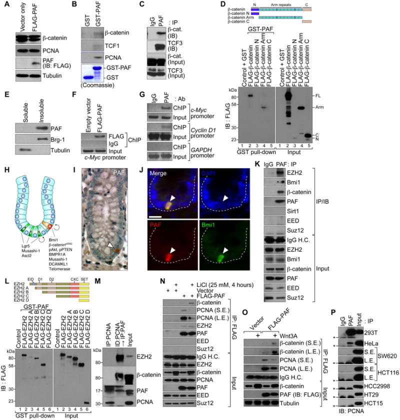
(A) No effect of PAF on β-catenin protein stability.
HeLa cells were transiently transfected with FLAG-PAF-pcDNA for IB.
(B and C) Interaction of PAF with β-catenin and TCF/LEFs.
GST-PAF was used for pull-down with SW620 cell lysates and IB (B). SW620 cells were analyzed for co-immunoprecipitation (co-IP) and IB (C).
(D) PAF-β-catenin interaction via the armadillo repeat domain.
GST-PAF was incubated with in vitro transcribed and translated FLAG-tagged β-catenin deletion mutants (N, N-term; Arm, Armadillo repeat domain; C, C-term) and analyzed using GST pull-down and IB.
(E) PAF is a chromatin-associated protein.
A chromatin-associated lysate (insoluble) and a soluble fraction of HCT116 were analyzed for IB. Brg-1: a chromatin fraction control.
(F and G) PAF occupies TBEs.
HCT116 stably expressing FLAG-PAF (F) and parental cells (G) were analyzed using ChIP. GAPDH promoter: a negative control.
(H) Lgr5 positive and Bmi1 positive ISCs are located in the crypts. ISCs divide into transit-amplifying (TA) cells and differentiate into IECs. Wnt/β-catenin signaling is highly active in crypts containing ISCs and TA cells.
(I) PAF expression in IECs of crypts.
A small intestine tissue was immunostained for PAF (arrowhead); hematoxylin: blue.
(J) PAF expression in Bmi1 positive ISCs.
Immunofluorescent staining of murine colon tissue samples for PAF and Bmi1. Scale bars = 20 μm.
(K) PAF interaction with EZH2.
Co-IP and IB assays of SW620. H.C.; heavy chain.
(L) PAF-EZH2 interaction via the CXC region.
GST-PAF was incubated with HeLa cell lysates expressing each FLAG-EZH2 deletion mutant (A-D), and analyzed for GST pull-down and IB. EID, EED interaction domain; D1 and D2, homologous domains 1 and 2; CXC, cysteine-rich domain; SET, SU(var)3-9, E(z), and Trithorax histone methyltransferase domain.
(M) Interaction of PAF with EZH2 and β-catenin, independently of PCNA. Co-IP of HCT116. A PCNA-immunodepleted (ID) supernatant was used for IP and IB.
(N and O) Wnt-dependent PAF-β-catenin interaction.
HeLa (vector or FLAG-PAF) were treated with LiCl (25 mM, 4 h) (N) or Wnt3A (100 ng/ml, 4 h) (O), and analyzed for co-IP and IB. L.E.: long exposure; S.E.: short exposure.
(P) PAF-PCNA interaction in colon cancer cells.
Co-IP and IB assays. Asterisks: IgG light chain.