
Figure 5. Induction of intestinal neoplasia by PAF expression.
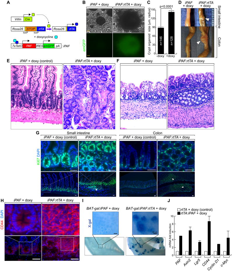
(A) PAF conditional inducible mouse models.
(B and C) Colonic crypts were isolated from Rosa26-rtTA:iPAF mice and maintained with or without doxy treatment (2 weeks). Phase-contrast images (B); quantification of the size of crypt organoids (C).
(D-F) Induction of PAF expression develops intestinal microadenoma.
Villin-Cre:Rosa26-LSL-rtTA:iPAF mice (experimental group) and iPAF mice (control) were given doxy (2 mg/ml in drinking water; small intestine [2 months] and colon [4 months]). Arrowheads: adenomas (D). H&E staining of small intestine (E) and colon (F). Dotted circles: aberrant crypt foci.
(G) IEC hyperproliferation by PAF. Villin-Cre:Rosa26-LSL-rtTA:iPAF and iPAF mice were given doxy (2 months). Ki67 immunostaining; arrowheads: hyperplastic lesions. Scale bars = 50 μm.
(H) Upregulation of CD44 by PAF.
CD44 immunostaining of small intestine specimens from control and PAF-induced (4 months) mice. Scale bars = 500 μm.
(I) PAF hyperactivates β-catenin reporter activity.
X-gal staining of iPAF:BAT-gal and Rosa26-rtTA:iPAF:BAT-gal mice (doxy [7 days]). Dotted circles: endogenous Wnt signaling activity in crypts.
(J) Upregulation of β-catenin target genes by PAF.
Crypts isolated from Rosa26-rtTA:iPAF mice were treated with doxy (1 μg/ml, 36 h) for qRT-PCR.
All error bars indicate standard deviation.