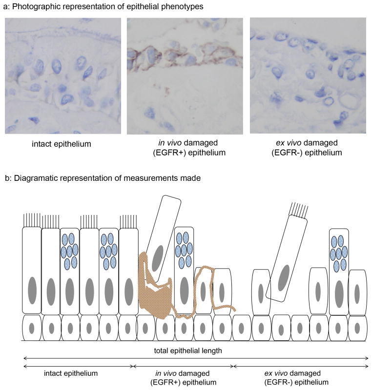
Figure 2. Assessment of epithelial integrity.
Staining for EGFR was used to distinguish epithelium that had sustained damage in vivo (EGFR+)(a: middle plate) from that that had been damaged during bronchoscopy or processing (EGFR−)(a: right hand plate). Intact undamaged epithelium was EGFR negative (a: left hand plate). The total epithelial length and the lengths of intact, in vivo and ex vivo damaged epithelium were measured using computer assisted image analysis (b).

