
Figure 5. Influx of inflammatory cells into the airway smooth muscle.
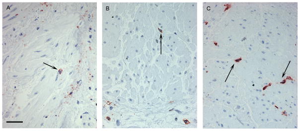
Immunohistochemical staining showing the influx of eosinophils (a), T cells (b) and mast cells (c) into the smooth muscle. Inflammatory cells are stained red and those within the ASM bundle were counted (↑). Scale bar is 50μm.