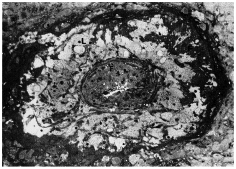
Figure 3. Atherosis.

Numerous lipid-laden cells (L) and fibrin deposition (F) are present in the media of this occluded decidual vessel(Reprinted from Sheppard B, Bonnar J. Uteroplacental arteries and hypertensive pregnancy. In: Bonnar J, MacGillivray I, Symonds G, editors. Pregnancy Hypertension. Baltimore: University Park; 1980. p. 205).52
