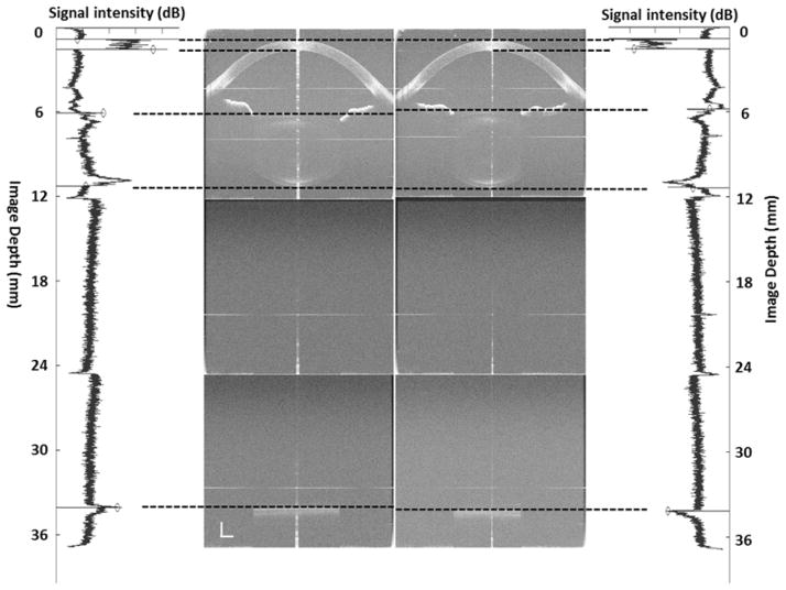
Figure 1.
The left side shows the optical length of whole eye imaging with ultra-long scan depth optical coherence tomography from a 25-year-old subject with myopia (−1.0 diopter) and the corresponding longitudinal reflectivity profiles produced by custom-developed software at the rest state. The right side shows the optical length of whole eye imaging with ultra-long scan depth optical coherence tomography from the same subject and the corresponding longitudinal reflectivity profiles produced by custom-developed software during accommodation (+6D). During accommodation, the axial biometry of the whole eye changes, including a decrease in ACD and VL, and an increase in LT and AL (paired t-test, p < 0.001). At the wavelength of 840 nm, the refractive indices of the cornea, aqueous humor, crystalline lens and vitreous were 1.387, 1.342, 1.408 and 1.341, respectively. Bars = 1 mm.
ACD: anterior chamber depth; LT: lens thickness; VL: vitreous length; AL: axial length.

