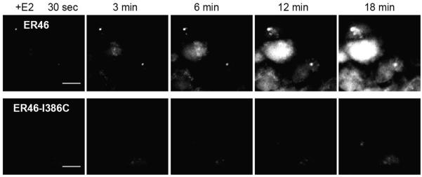
Figure 2.

NO detection in live COS-7 cells. COS-7 cells were transfected with plasmids encoding eNOS and either ER46 or ER46-Ile386Cys. Sequential imaging was performed at 37 °C after loading cells with DAF-FM diacetate. 30 nM E2 was added at 30 sec of imaging, with imaging time points indicated. Scale bar = 20 μm. Reproduced from Kim et al., (2011).
