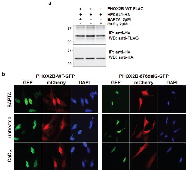
Figure 3.
The PHOX2B-HPCAL1 interaction is not calcium dependent. (a) PHOX2B-WT-FLAG and HPCAL1-HA constructs were transfected into 293T cells and harvested 48 hours later. Either BAPTA-AM [1,2-Bis(2-aminophenoxy)ethane-N,N,N′,N′-tetraacetic acid tetrakis (acetoxymethyl ester)] (3 μM) or CaCl2 (2 μM) was added to the cell lysates at the indicated concentrations, immunoprecipitated with anti-HA antibodies and analyzed by western blotting with anti-HA and anti-FLAG antibodies. (b) HPCAL1-mCherry and PHOX2B-GFP (both WT and the 676delG mutant) constructs were transiently transfected into HeLa cells and either BAPTA-AM or CaCl2 was added to the media at the indicated concentrations. Cells were fixed and imaged 2 days after transfection as described for Figure 2.

