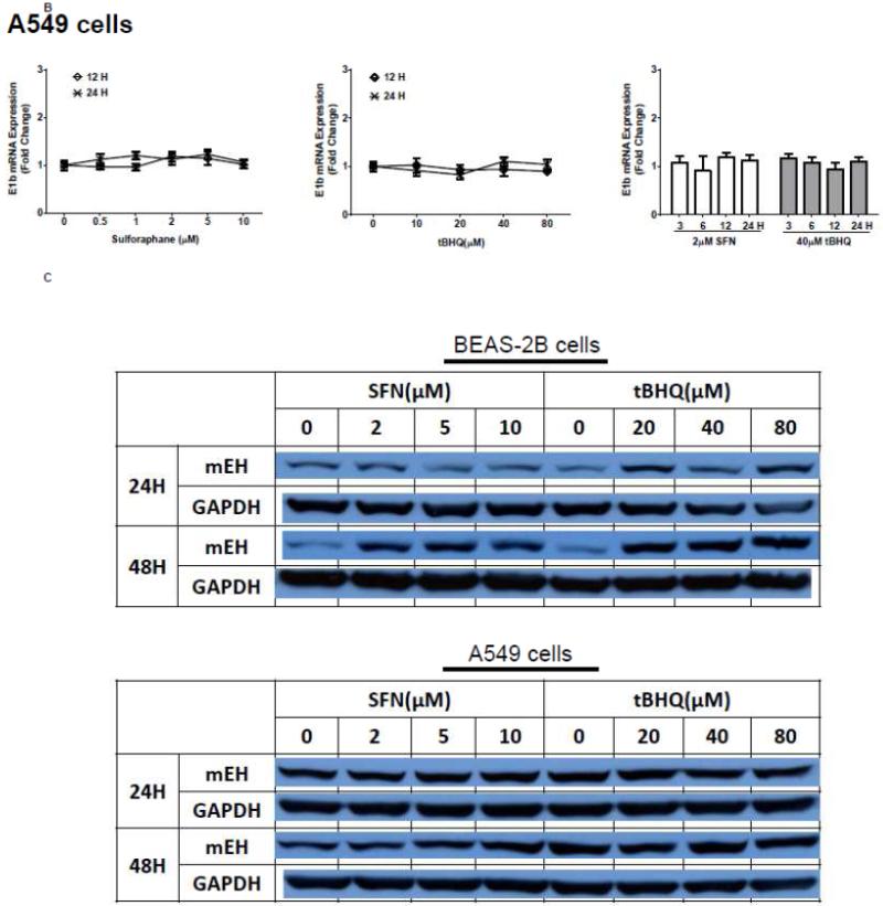
Figure 1. Antioxidants induce E1b expression.
A.) BEAS-2B cells and B.) A549 cells were treated with DMSO, sulforaphane or tBHQ for different time periods and E1b mRNA expression was analyzed by the real time quantitative PCR. GAPDH was used as an internal control. Results are expressed as the mean ± SD. Significant differences from DMSO control are indicated by “*” (p<0.05). C.) The induction of mEH protein in BEAS-2B and A549 cells was evaluated by western blotting. Whole cell lysates were immunoblotted to detect mEH protein. GAPDH was used as a loading control. All experiments were performed three times, and one representative set of data are showed.


