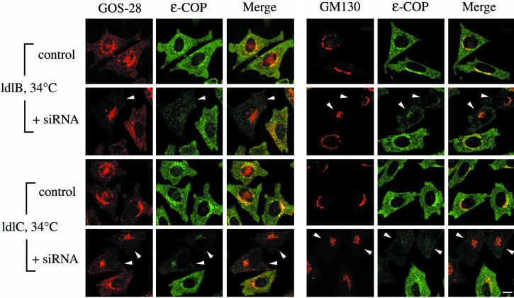
Figure 7.
Effects of siRNA depletion of ε-COP in ldlB and ldlC cells on the immunofluorescence of GOS-28, GM130, and ε-COP. ldlB and ldlC mutants were transfected without (control) or with (+siRNA) ε-COP–specific siRNA. Cells were incubated at 34°C for 48 h and then fixed and stained with antibodies to either GOS-28 (red) or GM130 (red) and ε-COP (green). To facilitate visualization of the distribution of GOS-28 by using confocal microscopy, the images from ldlB and ldlC cells (weaker signals) were collected with relatively high signal gains. Arrowheads indicate cells in which there was a loss or reduction the signal intensity for ε-COP. Bar, 10 μm.

