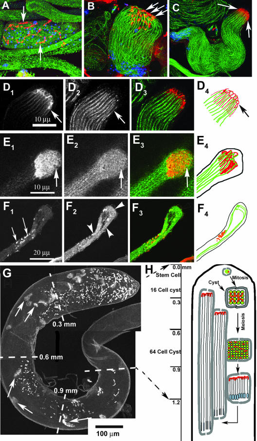
Figure 3.
Spectrin scaffold of fusomes is reorganized to form the cylindrical spectrin caps at the growing ends of axoneme. A–D show anti-β-spectrin (colored red) and anti-β-tubulin (colored green) staining in developing spermatids of wild-type testis. The nuclear DNA (colored blue) is stained using Sytox Green. (A) Postmeiotic stage cyst with regular organization of fusomes (arrow) and 64 nuclei inside. (B) Elongating stage cyst with the spermatid heads (blue) bundled together at one end and condensed spectrin staining (arrows) at the other end. The axonemal microtubules (green) are marked by anti-β-tubulin. (C) The β-spectrin staining (arrows) at the growing ends of axoneme seems compact at a later stage, and the spermatids also seemed uniformly bundled. (D1–4) Bundle of cortical spectrin-rich structures (arrows in D1) is visible at the distal end when the cyst is between 1.5 and 2 mm in length, and each one seems to cap individual axoneme (arrows in D2). A red green merge of D1 and D2 is presented in D3, and D4 shows a tracing of individual axoneme (green) and the spectrin caps at this stage. (E1–4) Distal end of a wild-type cyst stained with α-spectrin (E1) and α-tubulin (E2), respectively. Arrows indicate the distal end in E1–3 and E3 shows a red green merge of E1 and E2. A tracing of α-spectrin and α-tubulin staining is shown in E4. (F1–4) Similar staining in ddlc1ins1 cyst shows that the spectrin-rich honeycomb structure is deformed into punctate spots (F1, arrows) and abnormally twisted axonemes (F2, arrowheads). (G) Anti-α-spectrin staining of intact testis depicts a regular distribution of the spectrin scaffolds (arrows) along its length. These are called elongation cone (EC) in this study. (H) Illustration indicates the orientation and position of the spermatid heads (blue) and the spectrin scaffolds (red) within a cyst at different stages of spermatogenesis inside the testis.

