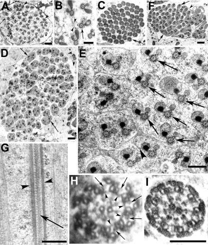
Figure 7.
Mutations in ddlc1 affects the cellularization of spermatids. Electron microscopic images of transverse sections of ddlc1rev (A–C), and ddlc1DIIA82 (D–F) testes collected from 2-d-old adults. (A) Section close to the caudal end of a cyst shows 64 axoneme-mitochondria sets mostly wrapped in individual axonemal sheath connected by thin membranous bridges (arrows). But often, more than one axoneme-mitochondria set was found within a single envelope (arrowheads). (B) Axoneme (arrow) and the major (M) and minor (m) mitochondrial derivatives surrounded by cellular microtubules are in syncytium at the caudal most end, and some incompletely formed membrane lamellae with vesicular bodies (arrowheads) attached were seen in this region. (C) Matured cysts showing individually wrapped axoneme-mitochondria pairs (arrow). (D) Mega cyst from ddlc1DIIA82 testis containing 112 axonemes and with multiple axonemes within a single envelope (arrows). In total, 64 such envelopes were visible in this section. (E) Higher magnification image of a part of a different cyst from the same section shows incompletely formed axonemal sheath (arrowhead) and multiple axoneme-mitochondria sets (arrows) within a single cellular envelope. The morphology of the axoneme-mitochondria complex seemed normal. (F) Section of a seemingly mature cyst from ddlc1DIIA82 testis showing incompletely individualized spermatids (arrows) with multiple axoneme-mitochondria sets within one envelope and interconnected by thin membranous bridges (arrowhead). (G) Longitudinal section through the axoneme from an immature (preindividualization) spermatid from ddlc1DIIA82 hemizygous testis revealed normal peripheral (arrowheads) and central (arrow) microtubule assembly. (H) Cross section of an axoneme at the growing end of the spermatid from the same sample revealed 9 + 2 organization of microtubules, the outer (arrows) and inner (arrowheads) Dynein arms, and the radial spoke structures. (I) Section of a relatively matured spermatid (postindividualization) from the mutant testis also showed normal organization. Bar, 1 μm in A, D, and F; 0.5 μm in B, E, and G; and 0.2 μm for H and I as shown in I.

