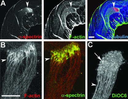
Figure 8.
Cortical F-actin assembly is formed around developing spermatids at the EC region. (A) Optical section of an intact adult testis stained with anti-α-spectrin (red), rhodamine isothiocyanate: phalloidin (green), and anti-α-tubulin (blue). Arrowheads indicate the distal end (EC) of a cyst and arrows indicate cortical localization of α-spectrin and F-actin around individual spermatids in the cyst. Note that the cortical F-actin as well as the axonemal α-tubulin staining end at the proximal part of the EC structure. (B) Distal end of an isolated cyst stained with phalloidin (red) and anti-α-spectrin (green). Arrowhead indicates cortical enrichment of F-actin and spectrin at the distal end of a single spermatid. (C) Single optical section of an isolated live cyst stained with DiOC6. Arrowhead indicates a single spermatid cell, and arrow indicates enrichment of staining in individual spermatids at the distal ends.

