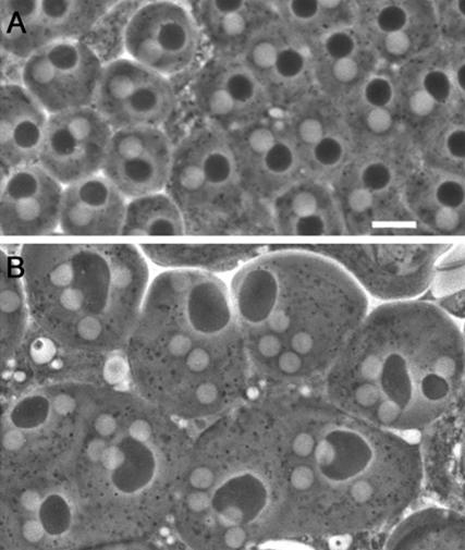
Figure 1.
Spermatid morphology in wild-type males and in cytokinesis-defective mutants. Spermatids are viewed by phase contrast microscopy. (A) Wild-type spermatids containing a single phase-light nucleus associated with a single phase-dark nebenkern of similar size. (B) Spermatids from funnel cakes (fun) mutants containing a large nebenkern associated with four or two nuclei of regular sizes. Bar, 10 μm.

