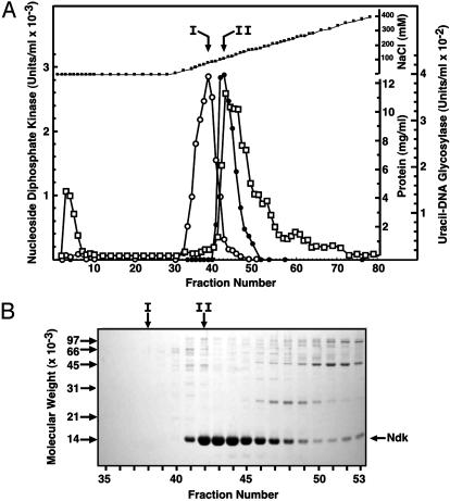
Fig. 2.
DEAE-Sepharose column elution profile of uracil-DNA glycosylase and Ndk activity. (A) DEAE-Sepharose column chromatography and standard assays for uracil-DNA glycosylase (open circles) and Ndk (filled circles) were performed as described in Materials and Methods. Protein concentration (open squares) and NaCl concentration (filled squares) were determined as described in Materials and Methods. (B) 12% SDS/PAGE of DEAE-Sepharose column fractions was conducted as described in Materials and Methods. The molecular weight standards are indicated by arrows on the left side of the gel; the location of the Ndk band is shown on the right side. Fractions corresponding to the peak of uracil-DNA glycosylase (I) and Ndk (II) activity are indicated by vertical arrows.

