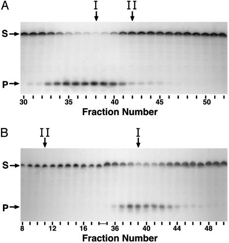
Fig. 4.
Detection of uracil-DNA glycosylase activity by using a uracil-containing oligodeoxynucleotide substrate. Reaction mixtures (40 μl) containing 2 pmol of 5′-end 32P-labeled duplex oligonucleotide ([32P]U·A 21-mer) and 10 μl of various DEAE-Sepharose (A) or hydroxyapatite (B) fractions were prepared and processed as described in Materials and Methods. Samples (10 μl) of the reaction products were resolved by using denaturing 12% polyacrylamide/8.3 M urea gel electrophoresis. Arrows indicate the location on the autoradiogram of unreacted [32P]21-mer substrate (S) and [32P]9-mer products (P). Fractions corresponding to the peak of Ung (I) and Ndk (II) activity are indicated by vertical arrows.

