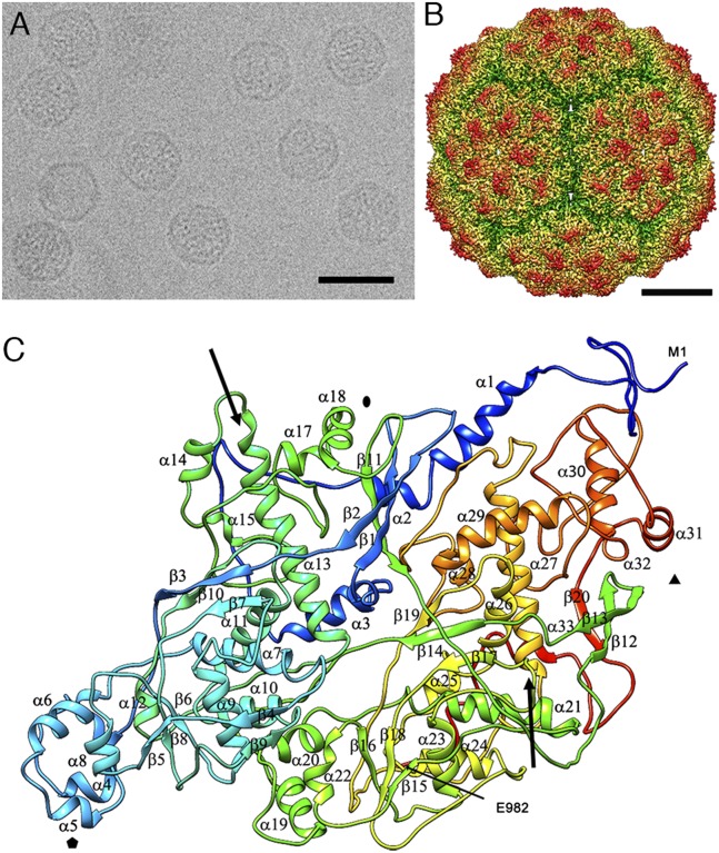
Fig. 1.
Three-dimensional cryo-EM reconstruction of PcV virions at a resolution of 4.1 Å. (A) Cryo-EM image of PcV. (Scale bar: 500 Å.) (B) Radially color-coded, surface-shaded virion capsid viewed along a twofold axis. Protruding pentamers (orange) are visible in the T = 1 lattice. (Scale bar: 100 Å.) (C) Ribbon diagram of the PcV capsid protein (top view), rainbow-colored from blue (N terminus) to red (C terminus). The first (Met1) and last (Glu982) residues are indicated. Symbols indicate icosahedral symmetry axes, and arrows indicate the longest helices: α13 and α27.

