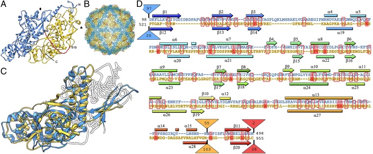
Fig. 2.
PcV capsid protein is a structural duplication. (A) Atomic model of a PcV CP showing the N-terminal domain A (1–498, blue), the linker segment (499–515, red), and the C-terminal domain B (516–982, yellow). (B) Atomic model of the PcV capsid viewed along a twofold axis. (C) Superimposed A and B domains (white regions indicate nonsuperimposed regions for both domains). (D) Sequence alignment of domains A (blue) and B (yellow) resulting from the Dali structural alignment. The α-helices (rectangles) and β-strands (arrows) are rainbow-colored from blue (N terminus) to red (C terminus) for each domain. Triangles represent nonaligned segments (sizes indicated). Strictly conserved residues are on a red background, and partially conserved residues are in a red rectangle.

