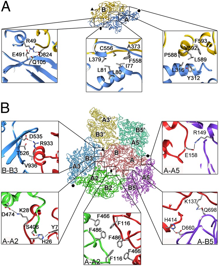
Fig. 3.
Intra- and intermolecular interactions in the PcV CP. (A) Intrasubunit interactions between PcV CP domains A (blue) and B (yellow) (top view). Close-up views show three regions of the interface between domains A and B. The interface near the twofold axis (oval) has mostly polar interactions (Left), whereas hydrophobic interactions dominate in the region closer to the fivefold axis (Center and Right) (Table S1). (B) Intersubunit interactions. A PcV CP subunit (red) with surrounding subunits (green, yellow, blue, light green, and violet) is shown; each CP interacts with seven domains of adjacent CP subunits, mediated by 110 residues. The symmetry relationships of domains A and B relative to the subunit labeled A-B (red) are indicated (e.g., domains A5 are symmetrically related by a fivefold axis to domain A). Close-up views show representative interfaces. A comprehensive list of interactions is given in Table S2.

