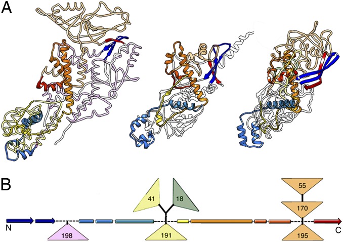
Fig. 6.
Structural comparison of PcV, L-A, and consensus reovirus CP. (A) Structural motifs common to the reovirus CP consensus model (Left), PcV A domain (Center), and L-A Gag (Right). Conserved SSEs are rainbow-colored (bright), and nonconserved segments or insertions at the N-terminal (pale pink), middle (pale yellow), or C-terminal (pale orange) region are also shown. (B) Scheme showing the conserved SSEs among dsRNA virus CPs; triangles indicate insertions (sizes are given), rectangles indicate α-helices, and arrows indicate β-strands.

