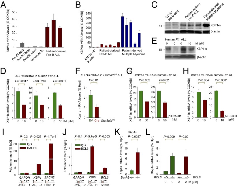
Fig. 2.
Regulation of XBP1 by STAT5, ERK, and AKT (positive) and BACH2 and BCL6 (negative) in Ph+ ALL. (A) XBP1-s mRNA levels were measured by qRT-PCR in sorted B-cell fractions from healthy human donors compared with patient-derived pre-B ALL cells (ALL-Q5, ALL-O2, ALL-X2, BLQ11, and LAX9; n = 3) and (B) in two CD19+ MACS-enriched pre–B-cell samples and five patient-derived multiple myeloma samples (MM1–5; n = 3). (C) Western blot analysis of spliced XBP1-s for MACS-enriched normal human CD19+ pre-B cells compared with patient-derived pre-B ALL cells (as in B) and one patient-derived multiple myeloma (MM6) sample using β-actin as loading control. (D) XBP1-s mRNA levels were measured in Ph+ ALL cell lines (NALM1, TOM1, and BV173) treated with or without the TKI IM for 16 h (10 µM IM) by qRT-PCR (n = 3). (E) Protein levels of XBP1-s were measured by Western blot analysis in patient-derived Ph+ ALL cases (LAX9 and PDX59) treated with IM for 16 h (10 µM IM) using β-actin as loading control. (F) Xbp1-s mRNA levels were measured in Stat5a/bfl/fl ALL cells with EV control and 4-OHT–inducible Cre (Cre) after 24 h by qRT-PCR. (G and H) XBP1-s mRNA levels were measured in patient-derived pre-B ALL cases (PDX2 and LAX7R) treated with or without PD325901 or AZD5363 for 5 h (50 nM PD325901; 10 µM AZD5363) by qRT-PCR (n = 3). (I) Quantitative ChIP validation of BACH2 binding to the XBP1 promoter in a human Ph+ ALL patient sample (ICN1), GAPDH as a negative and BACH2 as a positive control.(J) Quantitative ChIP validation of BCL6 binding to the XBP1 promoter in human pre-B ALL cells. (K) XBP1 expression was assessed in Bach2−/− ALL cells compared with WT controls (Bach2+/+) by qRT-PCR using specific primers for spliced Xbp1-s (n = 3). (L) Xbp1s mRNA levels were measured by qRT-PCR in Bcl6−/− ALL cells compared with WT controls (Bcl6+/+) treated either with or without the TKI IM for 16 h (2 µM IM).

