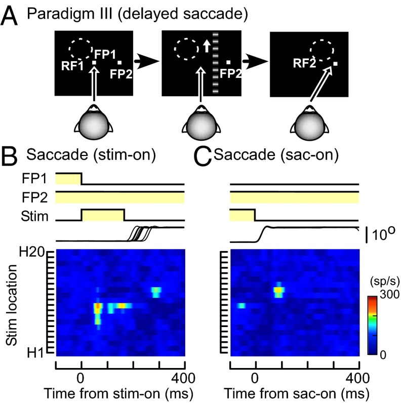
Fig. 3.
Responses of the MST neuron during delayed saccades (paradigm III). (A) Schematic diagram showing the sequence of paradigm III. The saccade target (FP2) was turned on 300 ms before onset of the moving grating and offset of the fixation point (FP1). The moving grating was turned off before the saccade onset, as in paradigm II. (B and C) Spatiotemporal RF maps of the MST neuron shown in Figs. 1 and 2 across the period of saccades (B and C). Responses are aligned at stimulus onset (B) or saccade onset (C). RF map, stimulus, and eye traces are as in Fig. 1.

