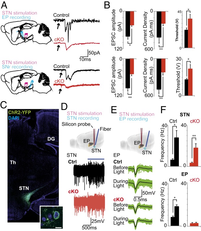
Fig. 2.
Attenuation of Vglut2 expression in the STN severely impairs synaptic communication postsynaptically. (A) (Left) Diagram showing stimulation and recording electrode placement in parasagittal slices containing the STN, EP, and SNr. (Right) Representative examples of synaptic currents elicited by STN stimulation (10% above threshold) in SNr and EP cells in control and cKO mice. (B) Mean EPSC amplitude, current density, and stimulation threshold for EP and SNr cells in control (black bars) and cKO (red bars) mice. (C) Photomicrograph showing representative Pitx2-Cre–driven ChR2 expression (through its reporting protein YFP) selectively in the STN with high magnification in inset. Dentate gyrus (DG) and thalamus (Th) are indicated for reference. (D) Example of extracellular recordings in the STN of control and cKO mice before and during light stimulation. (E) Single units isolated from EP recordings before and during light stimulation. (F) Summary of firing frequency in STN and EP recordings before and during light stimulation in control (black bars) and cKO (red bars) mice. *P < 0.05; **P < 0.001. cKO, conditional knockout; Ctrl, control.

