
Fig. 7.
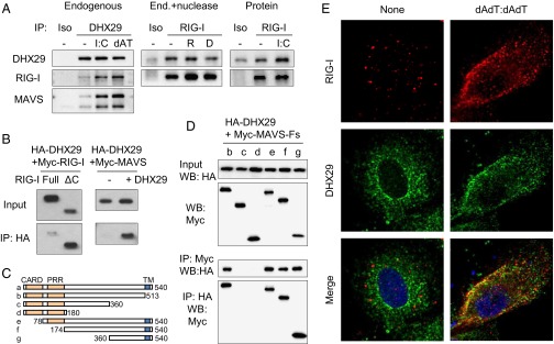
DHX29 interacts with RIG-I and MAVS. (A, Left) MRC5 cells stimulated with or without poly I:C or poly dAdT:dAdT (dAT) were lysed and mixed with anti-DHX29 antibody or with an isotype control and then incubated with Protein G agarose. Immunoprecipitated proteins were detected by Western blotting using anti-DHX29, anti–RIG-I, and anti-MAVS antibodies. (Center) Poly I:C-stimulated MRC5 cell lysates were treated with or without RNase (R) or DNase (D) before immunoprecipitation with anti–RIG-I antibody or with an isotype control. (Right) Highly purified DHX29 and RIG-I proteins with or without poly I:C were immunoprecipitated with anti-RIG-I antibody or with an isotype control. (B) Cell lysates from 293T cells overexpressed with Myc-tagged RIG-I (Myc–RIG-I; full-length or C-terminal deleted form) and HA-tagged DHX29 (Left), or with Myc-tagged MAVS (Myc-MAVS) with or without HA-DHX29 (Right), were incubated with anti-HA agarose beads. After 2 h, the beads were washed thoroughly and their binding proteins were eluted. The inputs and immunoprecipitated elutes (IP) were detected by Western blotting analysis using anti-Myc antibody. (C and D) Cell lysates from 293T cells overexpressed with HA-DHX29 together with various deletion mutants of Myc-MAVS (Myc-MAVS-Fs) were incubated with anti-HA or anti-Myc agarose beads. After 2 h, the beads were subjected to the same procedure as in B and were detected using anti-HA or anti-Myc antibodies. Data are representative of at least three independent experiments. (E) A549 cells were treated with or without poly dAdT:dAdT stimulation. After 6 h, the cells were fixed and stained with anti–RIG-I and anti-DHX29 antibodies and were observed with confocal microscopy. Red, RIG-I; green, DHX29; blue, DAPI.