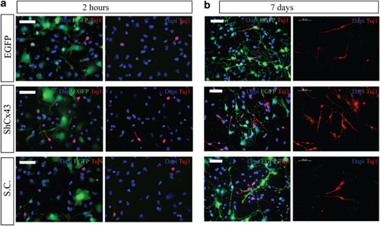
Figure 4.
Tuj1 expression in viral transduced hNPCs and differentiated for 2 h or 7 days. Fluorescence microscopy of viral transduced hNPCs 2 h (a) and 7 days (b) differentiated cultures. Left panels represent EGFP protein (green) expressing cells stained for Tuj1 (red). Cell nuclei are indicated by DAPI (blue) staining. Right panels indicate Tuj1 (red) and DAPI (blue) staining only. Scale bar 50 μm

