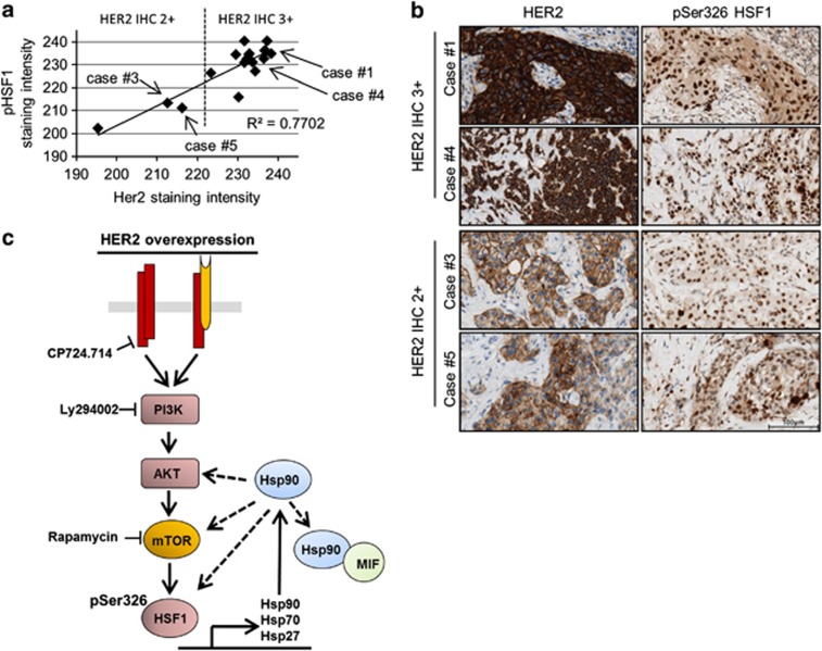
Figure 6.
In human HER2-positive breast cancers, HER2 levels correlate with pSer326 HSF1 activity. (a and b) Correlation in staining intensity between HER2 and activated HSF1 (pSer326) in human breast cancer. (a) Quantitative immunohistochemistry of HER2 and pSer326 HSF1 protein in HER2-amplified invasive ductal carcinoma with 2+ and 3+ HER2 staining intensity, respectively. Two regions encompassing >60 cells were quantified per case; DAB color intensity displayed as values on an inverted 8 bit gray scale, 0=white, 255=black. (b) Representative photomicrographs of cases of invasive ductal carcinoma quantified in a. Immunohistochemical staining for HER2 and pSer326 HSF1. DAB with hematoxylin counterstain, × 400 magnification. Bar, 100 μm. (c) Proposed model summarizing the findings of this study. In HER2-overexpressing cells, the PI3K–AKT–mTOR pathway is the main signaling axis that leads to phosphorylation of HSF1 at Ser326, which activates HSF1 transcriptional activity and induces, among other target genes, expression of heat-shock proteins. The activated HSP90 machinery stabilizes a broad panel of oncogenic and tumor-promoting proteins. Dashed lines mean feedback loop

