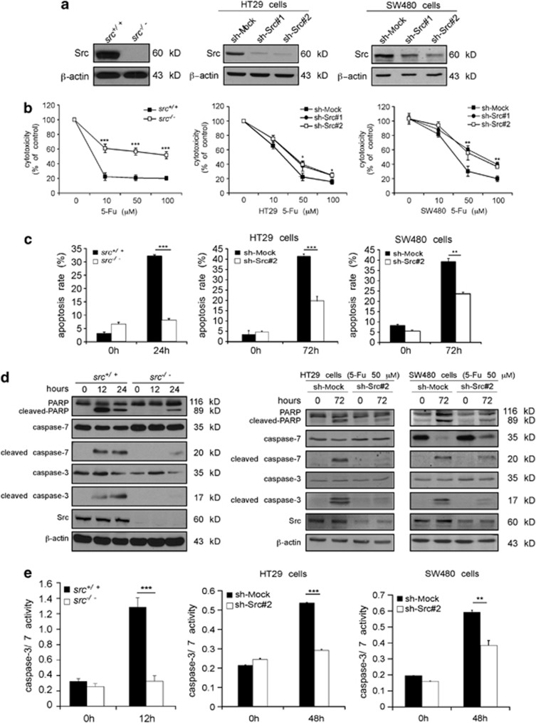
Figure 3.
Src contributes to 5-Fu-induced apoptosis. (a) The protein level of Src in wild-type (src+/+) and knockout (src−/−) MEFs was determined by western blotting. Sh-Mock and Src knockdown (sh-Src) cells were generated by stable infection with an sh-mock, sh-src#1 or sh-src#2 plasmid into HT29 and SW480 cells. Src protein levels were determined by western blotting. β-Actin was detected in the same membrane and served as a loading control. (b) Comparison of the cytotoxicity of 5-Fu against src+/+ and src−/− MEFs, or in sh-Mock- and sh-Src#1- or sh-Src#2-expressing cells. An MTS assay was used to assess cytotoxicity at 24 h for MEFs or 48 h for colon cancer cells after treatment with 5-Fu. Absorbance was read at 492 nm and data are expressed as percentage of untreated control (100%). Data are shown as means±S.D. of triplicate measurements. The asterisks indicate significantly (*P<0.05, **P<0.01, ***P<0.001) less cytotoxicity induced by 5-Fu. (c) 5-Fu-induced apoptosis was determined by flow cytometry. Data are shown as means±S.D. of triplicate measurements. The asterisks indicate significantly less apoptosis induced by 5-Fu (**P<0.01, ***P<0.001). (d) 5-Fu induces less cleaved PARP, cleaved caspase-7 and cleaved caspase-3 in cells expressing low levels of Src. 5-Fu (50 μM) was used to simulate MEFs and colon cancer cells. Total and cleaved PARP, caspase-7 and caspase-3 were detected by western blot analysis using specific antibodies. Data shown are representative of results from triplicate independent experiments. (e) Caspase-3/7 activity is decreased in cells expressing low levels of Src compared with cells expressing normal levels of Src after 5-Fu treatment. 5-Fu (50 μM) was used to simulate cells. After 12 or 48 h, cells were harvested, and caspase-3/7 activity was detected as described in ‘Materials and Methods'. Data are shown as means±S.D. of triplicate measurements. The asterisks indicate significantly lower caspase 3/7 activity (**P<0.01, ***P<0.001)

