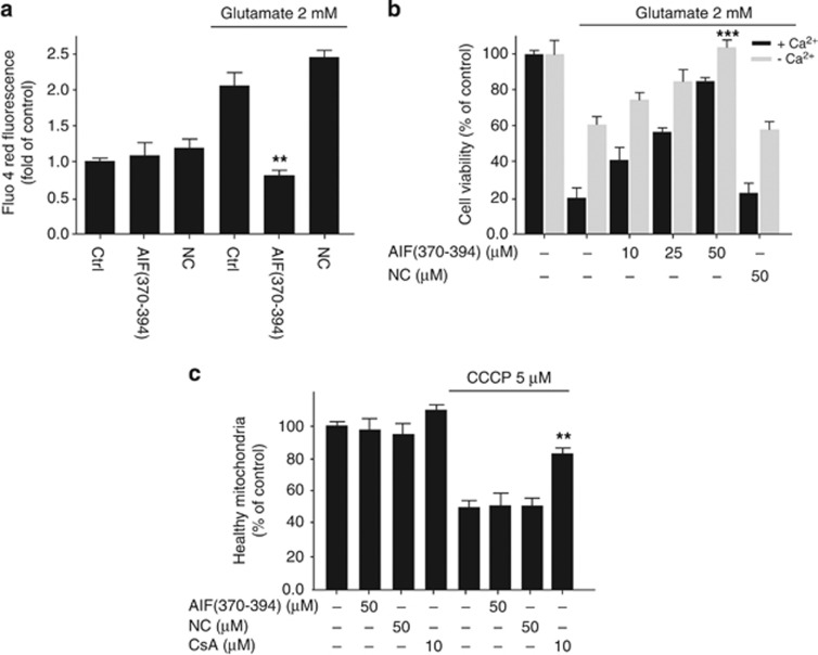
Figure 7.
CypA-siRNA and AIF(370–394) peptide block the oxidative stress-induced increase of intracellular Ca2+ concentration. (a and b) HT-22 cells were treated with 2 mM glutamate for 12–14 h. The intracellular concentration of Ca2+ was assessed by the fluorescent Ca2+ indicator Fluo-4 AM in 96-well plates. (b) Cell viability assessment of HT-22 cells received AIF(370–394) peptide at the indicated concentrations, followed by exposure to 2 mM glutamate for 12–14 h, either in a standard culture medium (black bars) or in a medium without Ca2+ (gray bars) (n=4, ***P<0.001). (c) Mitochondrial membrane depolarization induced with 50 μM CCCP in isolated mitochondria from HT-22 cells. Equal amounts of pure mitochondria were incubated or not (Ctrl) with peptides at concentrations of 50 μM and then treated with CCCP. CsA at a concentration of 10 μM was used as a positive control. Data show that AIF(370–394) as well as NC, differently to CsA, did not prevent the CCCP-mediated mitochondrial depolarization (n=4, **P<0.01)

