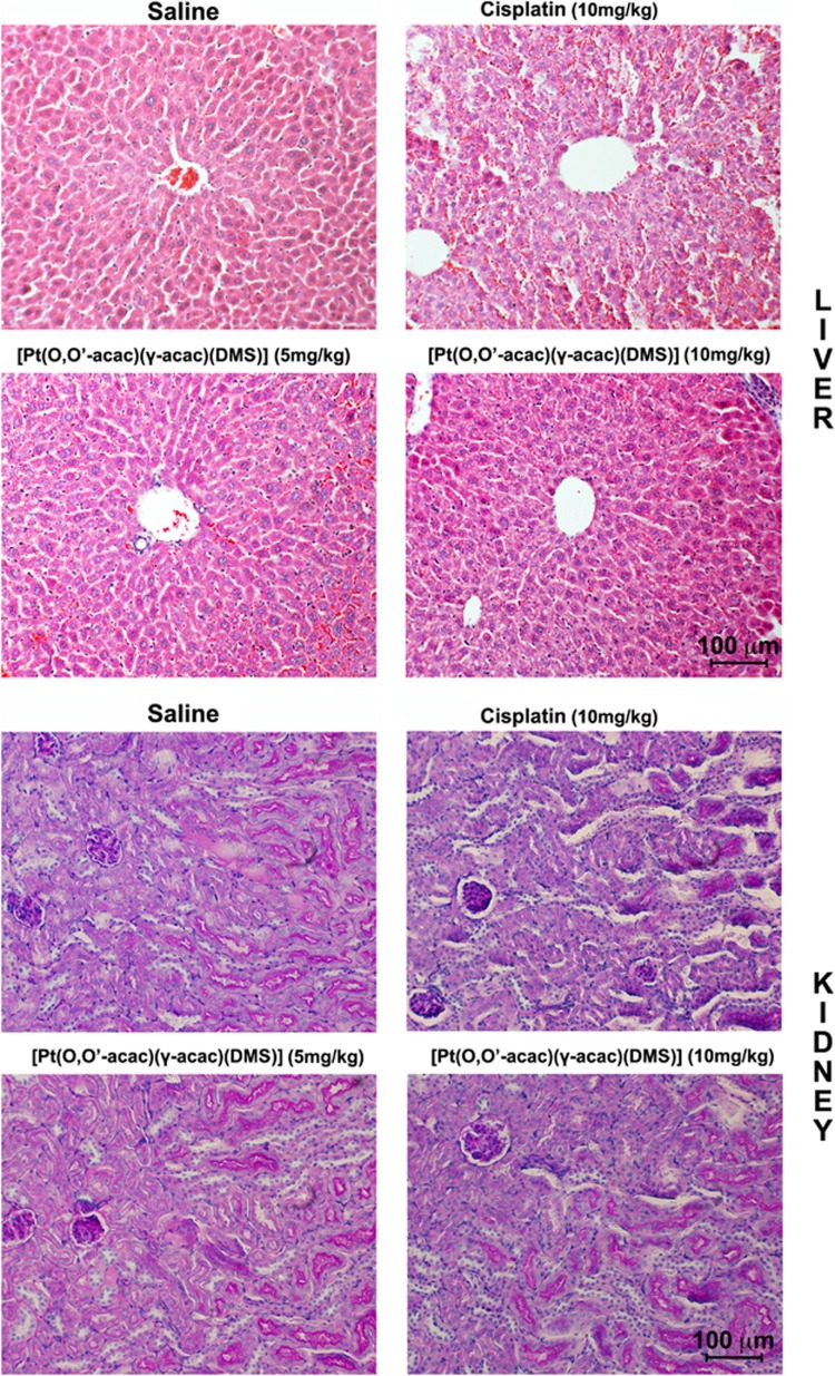
Figure 4.
Histopathological changes in liver and kidney tissues from toxicity analysis in female Balb/c mice. Light microscopy images of hematoxylin and eosin-stained liver (top panel, day 28) and kidney (down panel, day 28) were taken at a magnification of x20. Tissue samples were collected at day 28 following intravenous administration of [Pt(O,O'-acac)(γ-acac)(DMS)] or cisplatin at different doses. Scale bar 100 μm

