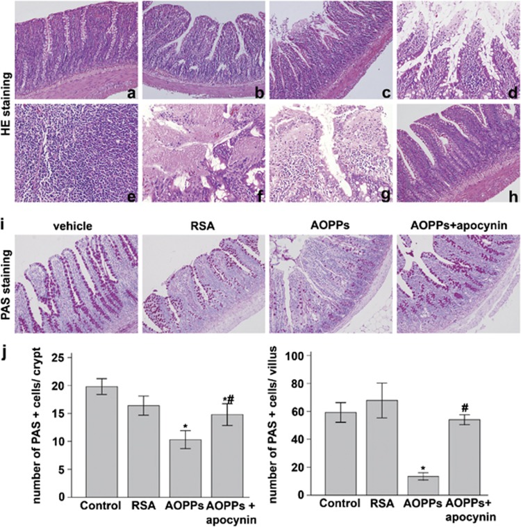
Figure 8.
AOPPs treatment of rats induced morphological changes of the small intestinal epithelium and altered the number of goblet cells. H&E staining showed almost normal intestine in (a) vehicle and (b) RSA groups, whereas (c, d) epithelial erosion and inflammatory cell invasion into the lamina propria and submucosal layer, (e) lymphoid follicle hyperplasia, (f) epithelial necrosis, and (g) epithelial exfoliation were found in AOPP-treated group. (h) Apocynin attenuated the degree of AOPP-induced tissue injury. (i) PAS staining in the small intestines of rats treated with or without AOPPs. (j) Quantification of goblet cells per crypt±S.D. of control, RSA, AOPPs, and AOPPs+apocynin group (n=6 per group). *P<0.05 versus control. #P<0.05 versus AOPPs

