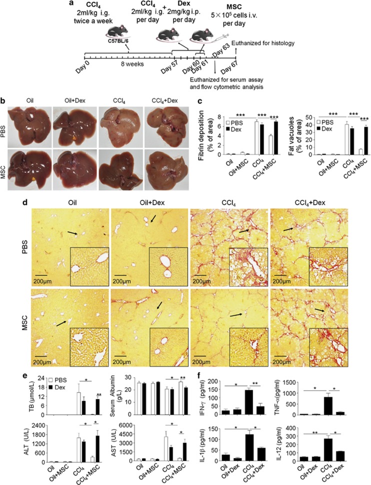
Figure 3.
Steroid co-administration abolished the therapeutic effects of MSCs on advanced liver fibrosis in mice. (a) Timeline of the protocol to induce liver fibrosis in mice, and treatment with Dex and MSCs. (b) On euthanization, the livers were collected and representative specimens from each group were photographed. (c) Quantitative analysis of presence of liver fibrosis and fat vacuoles. (d) Fibrin deposition was detected by sirius red staining ( × 40 magnification, scale bar=200 μm; arrows indicate portal areas, which were shown in lower right corner). The liver showed extensive fibrotic development, formation of regenerative nodules, and distortion of the vascular architecture in mouse cirrhotic liver without MSCs transfusion. (e) The serum concentrations of TB, Alb, ALT, and AST in mice from each group were determined. (f) On day 60 (after liver fibrosis induction with or without Dex injection), serum levels of IFN-γ, TNF-α, IL-1β, and IL-12 were assayed by the Luminex technology (Bio-Plex, Bio-Rad). For this figure, values are shown as mean±S.E.M. and statistical significance indicated as *P<0.05, **P<0.01, and ***P<0.001. Representative of three independent experiments; n=7–10

