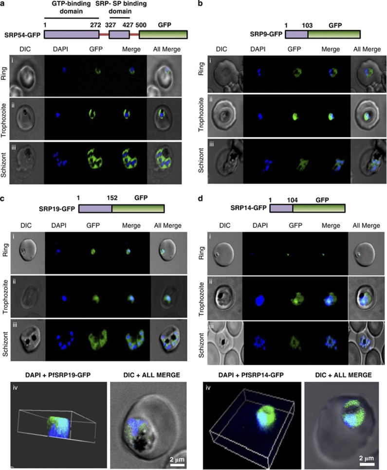
Figure 2.
Localisation of GFP fused PfSRP54, PfSRP19, PfSRP14 and PfSRP9 polypeptides. Upper panels of a, b, c and d show schematic representation of respective GFP fused PfSRP polypeptides. Lower panels show the expression of PfSRP polypeptides in three asexual blood stages of P. falciparum i.e. ring, trophozoite and schizont stage. Panel c(iv) and d(iv) show three dimensional reconstruction of confocal Z-stack merged images of GFP fused PfSRP19 and PfSRP14 (green), respectively, along with nuclear stain DAPI (blue)

