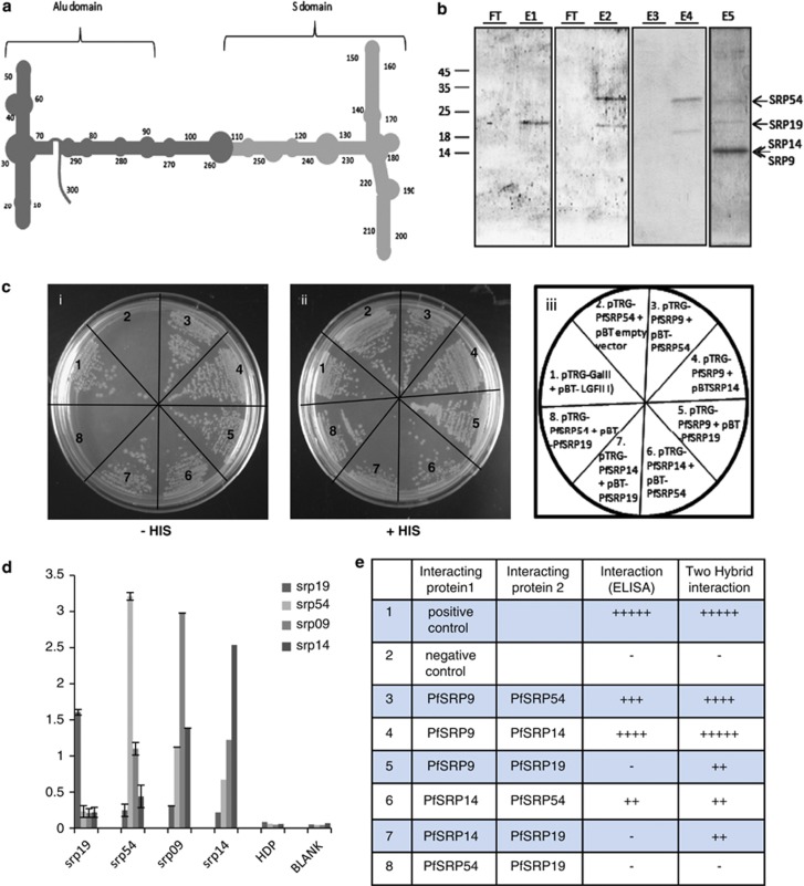
Figure 4.
Interaction between PfSRP polypeptides and SRPRNA. (a) schematic diagram of secondary structure of PfSRP RNA showing the Alu domain and S domain (b). Binding of SRP RNA to SRP 19 and M domain of SRP 54. FT represents the lanes with flow through. Elutes of S domain of SRP RNA binding (by DEAE sepharose method) showing the presence of SRP19 (E1), SRP19 and M-domain of SRP54( E2). Elutes of full SRP RNA binding (by DEAE sepharose method) with non-SRP protein PfHDP (E3) and PfSRP19 and SRP54 (E4), and all four PfSRP54 (M-domain), PfSRP19, PfSRP14, PfSRP9 (E5). (c) Interaction between protein components of PfSRP polypeptides using Bacterial Two Hybrid system. Growth of cotransformed reporter strain XL1-blue in dual selective (i) and non-selective (ii) medium, (iii) shows the plasmid constructs used to cotransform the reporter strain. (d) Interaction between protein components of PfSRP polypeptides using ELISA. X-axis shows the coated protein and Y-axis is absorbance read at 490 nm. (e) Table showing summary of interactions among all PfSRP polypeptides using both ELISA and Bacterial Two Hybrid system

