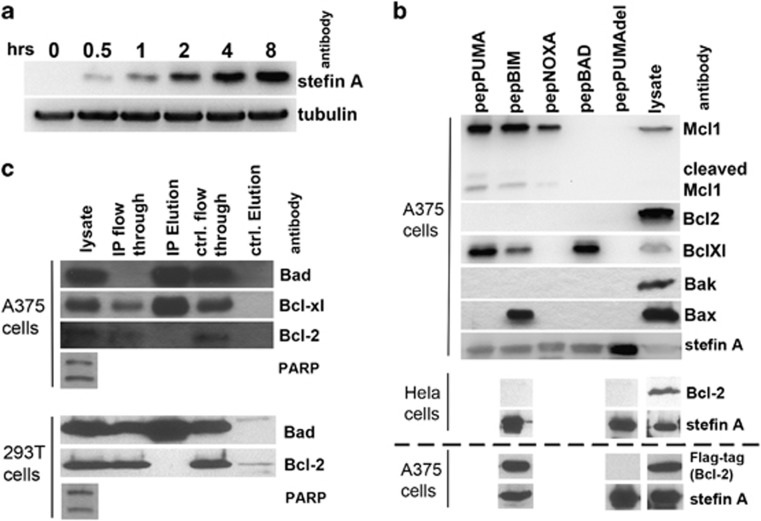
Figure 2.
PepBH3 expression and identification of their binding partners. (a) Following induction, pepBH3 expression is detectable after 30 min. (b) Co-IP of pro- and anti-apoptotic Bcl-2 proteins with pepBH3 following their expression in A375 (top panel) and Hela cells (middle panel). There is no interaction between Bcl-2 and pepBim in A375 or Hela cells, however, when Bcl-2 is overexpressed (in A375) binding to pepBim is significant (bottom panel, underneath dashed line). (c) IP of endogenous full-length Bad from A375 cell lysate reveals binding of Bcl-xl, but not Bcl-2 (top panel). Equally in 293T cells, the Bad:Bcl-2 interaction does not occur (bottom panel). PARP cleavage indicates that apoptosis was induced successfully in these cells

