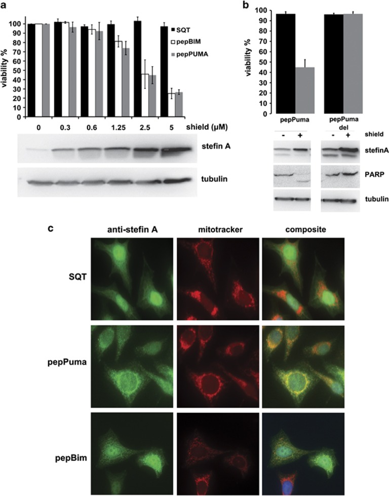
Figure 4.
Cell viability and localisation of pepPuma/Bim in cells. (a) PepPuma/Bim expression in A375 cells reduces cell viability by approximately 80%. The western blot shows levels of SQT with increasing amounts of Shield (error bars are ±S.D.; n=3). (b) PepPumaDel does not cause cell death (error bars are ±S.D.; n=3). (c) Immunofluorescence of Hela cells expressing SQT reveals no specific localisation pattern. PepBim or pepPuma (green) colocalise with mitotracker (red) in some but not all cells. DAPI-stained nuclei are shown in blue

